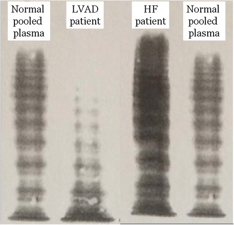
Fig. 2.

vWF multimer analysis in a low resolution gel (1.2 % SDS-agarose). The large multimers are found in the upper part of the gel. Results obtained from normal pooled plasma are compared to those from the LVAD recipients with an AvWS (presented in the Discussion section) and a HF patient with normal vWF multimers
