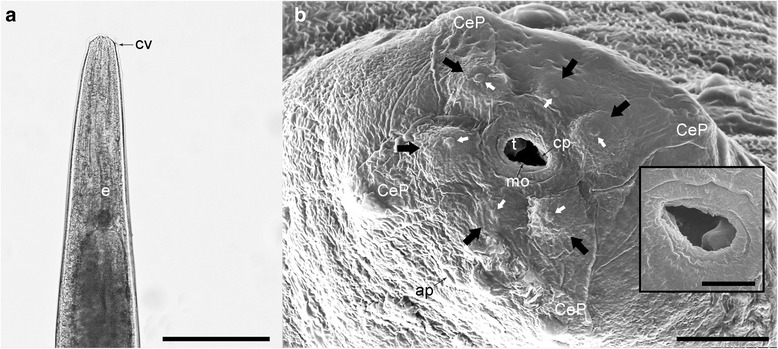
Fig. 3.

Light microscopy (a) and SEM photomicrographs (b) of the anterior extremity of A. daskalovi: The tooth and the cutting plate are clearly visible in the inset. Six labial papillae (black arrows) and six small protuberances (white arrows) are indicated. Abbreviations: ap, amphidial pore; CeP, four cephalic papillae; cp, cutting plate; cv, small cephalic vesicle; e, oesophagus; mo, mouth opening; t, tooth. Scale-bars: a, 500 μm; b, 10 μm; inset 5 μm
