
Fig. 1.
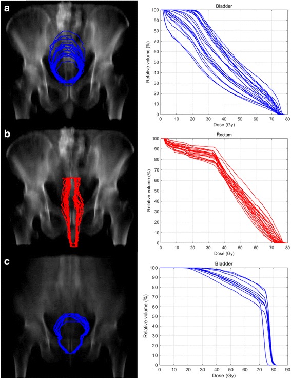
DRR of two patients. a–b show bladder and rectum contours for one patient derived from the different CBCTs projected on one DRR. The corresponded DVHs are displayed as well. c demonstrates different projections of the bladder contours on one DRR for a patient who had only very slight changed in his bladder filling