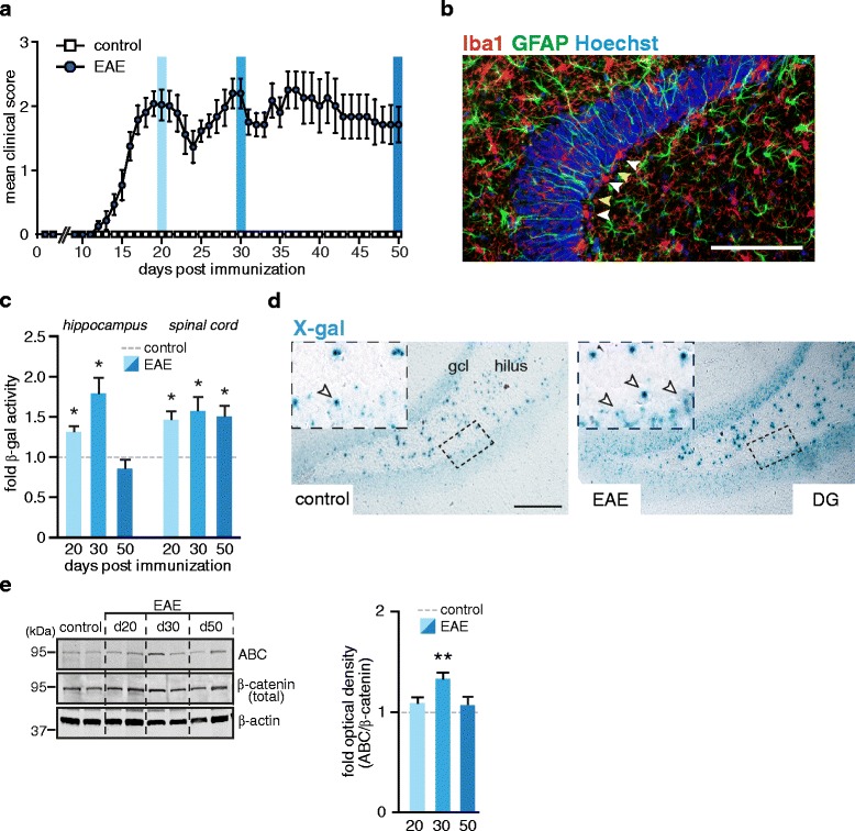
Fig. 1.

Analysis of Wnt activity in EAE. a Disease course in Axin2lacZ/+ mice with chronic EAE. EAE was induced by immunization with MOG35–55. Control group (CFA) were injected with an adjuvant cocktail without antigen. Data are shown as a mean clinical score ± SEM. Blue columns indicate the time points when mice were sacrificed for analysis: days 20, 30 and 50 post immunization. b Representative image of Iba1, GFAP co-immunostaining in hippocampal section of an EAE mouse at day 30. White arrow heads indicate reactive microglia Iba1+ (red) located in close proximity to radially oriented GFAP+ cells (green; yellow arrow heads) resembling radial glia progenitor cells of the SGZ. Nuclei were counterstained with Hoechst (blue). Scale bar: 100 μm. c β-gal assay of hippocampal and spinal cord tissue. Histogram represents mean + SEM of fold-changes of β-gal activity in EAE mice relative to respective controls (CFA) set as 1. Day 20 (EAE, n = 6; CFA, n = 3); day 30 (EAE, n = 4; CFA, n = 3); day 50 (EAE, n = 8; CFA, n = 4). Two-tailed, unpaired Student’s t-test, *p < 0.05. d Upregulation of Axin2 expression in the DG of EAE mice. Expression of lacZ and thus activation of Axin2 promoter were detected by X-gal staining on hippocampal sections from EAE mice (day 20) and respective controls (CFA). Representative images are shown. Top insets show magnified cells of the outlined SGZ region. Arrow heads mark X-gal positive cells. Gcl, granular cell layer. Scale bar, 100 μm. e Immunoblot analysis of β-catenin in the hippocampus of EAE and control (CFA) animals. Hippocampal proteins were extracted and processed for Western blotting with antibodies against active β-catenin (ABC) and total β-catenin. β-actin was probed for analysis of protein loading. Right histogram represents mean + SEM of fold-changes of normalized optical densities in EAE mice relative to controls (CFA) set as 1. Day 20 (n = 6); day 30 (n = 4); day 50 (n = 6) and control CFA mice (n = 6). Two-tailed, unpaired Student’s t-test, *p < 0.05 and ** p < 0.01
