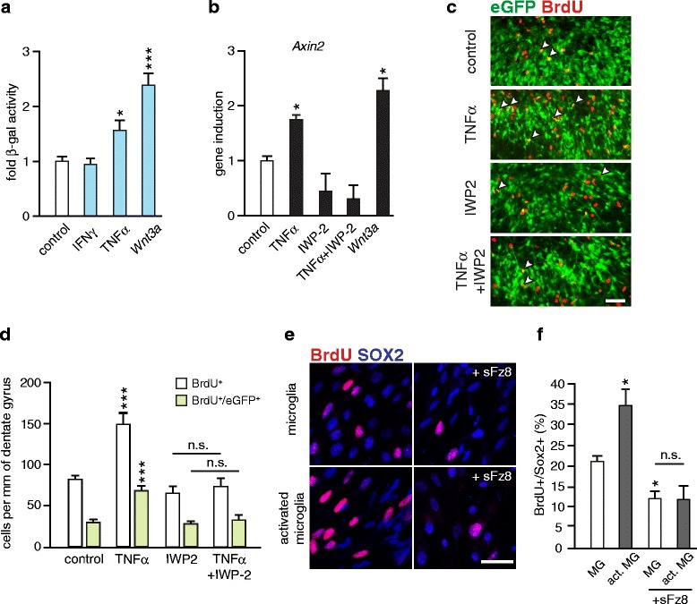
Fig. 5.

Analysis of Wnt activity in organotypic slice cultures (OSCs). a β-gal assay. Hippocampal Axin2lacZ/+ OSCs were treated for 6 h with Wnt3a (20 ng/ml), TNFα (1 ng/ml) or IFNγ (100 U/ml) and the medium was replaced with fresh cytokine-free medium for additional 24 h. Histogram represents mean + SEM of fold-changes of β-gal activity after treatment with Wnt3a (n = 15), TNFα (n = 14) or IFNγ (n = 14) relative to controls (PBS, n = 20) set as 1. Two-tailed, unpaired Student’s t-test, * p < 0.05 and *** p < 0.001. b qPCR analysis of the Axin2 gene expression. OSCs were treated as described above. IWP-2 (4 μM) was present in the media during the whole experiment. Histogram represents mean + SEM of fold-changes in Axin2 gene expression after treatment with Wnt3a (n = 3), TNFα (n = 4) or TNFα with IWP-2 (n = 3) relative to control (PBS, n = 4) OSCs set as 1. Gapdh was used as an endogenous reference. Two-tailed, unpaired Student’s t-test, * p < 0.05. c-d Histological analysis of proliferating cells in the DG of hippocampal NestineGFP OSCs. BrdU was administered to slice culture for 24 h prior to treatment with cytokines (performed as described for Fig. 5a-b). c Representative images show the immunostaining for BrdU (red) and eGFP (green). Arrow heads indicate BrdU+/eGFP+ co-labeled hippocampal progenitors. Scale bar, 50 μm. d Inhibition of Wnts secretion with IWP-2 (4 μm) abrogates the proliferative effect of TNFα on mitotic activity of hippocampal eGFP+ progenitors. Data are shown as mean + SEM of BrdU+ or BrdU+/eGFP+ co-labeled cells per mm of the DG. Control (PBS, n = 20 slices); TNFα (n = 9 slices), IFNγ (n = 11 slices) and Wnt3a (n = 9 slices); Two-tailed, unpaired Student’s t-test, * p < 0.05, ** p < 0.01 and *** p < 0.001. e-f TNFα - activated hippocampal microglia promotes the proliferation of hippocampal progenitors in vitro. Microglia cultured on transwell inserts were primed by TNFα (1 ng/ml) for 4 h. Transwells were set onto the hippocampal progenitor cultures in proliferating medium containing 5 ng/ml of bFGF in the presence or absence of soluble Frizzled-8 (Fz8; 1 μg/ml). After 24 h dividing cells were labelled with BrdU (2 h) and cells were fixed for immunocytochemistry. e Representative images of SOX2+ (blue) hippocampal progenitors stained for BrdU (red). Scale bar, 25 μm. f Quantification of BrdU-retaining SOX2+ cells. Data are shown as mean of four experiments + SEM. Two-tailed, unpaired Student’s t-test,* p < 0.05
