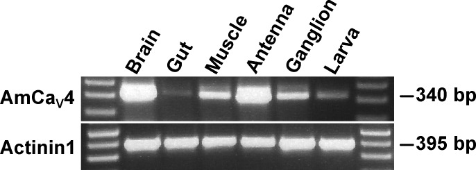
Figure 2.
Tissue expression of CaV4 in the honeybee. The tissue-specific expression of AmCaV4 was assessed by RT-PCR. All the honeybee tissue samples were processed using the same preparation steps, from dissection to gel electrophoresis. The expected weights of the amplicons are given in base pairs. Actinin1 was used as a positive control.

