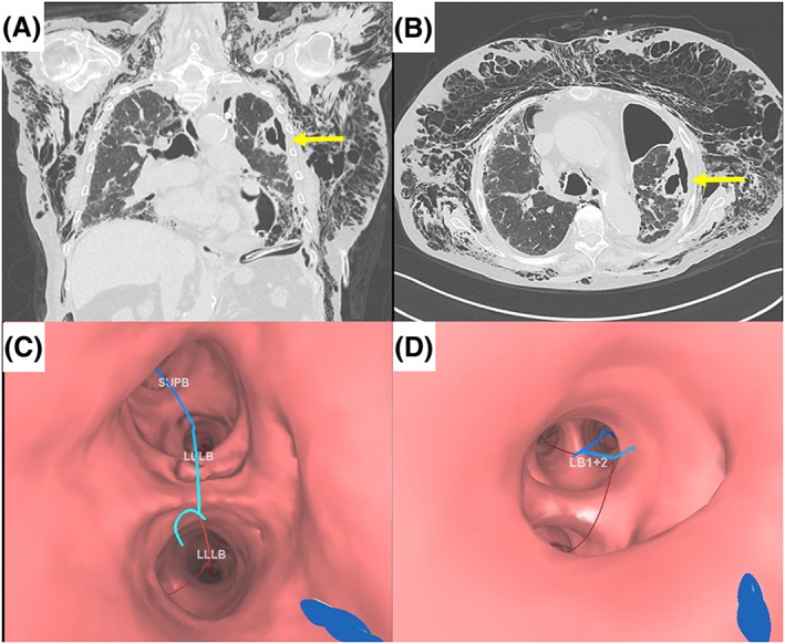
Figure 1.

(A, B) Chest computed tomography images on day 48 show severe pneumoderma and a cavitary lesion with a pinhole on the left S1 + 2 (arrows). (C, D) Virtual bronchoscopic navigation images on LungPoint® indicate a bronchial route to the left B1 + 2a to the cavitary lesion.
