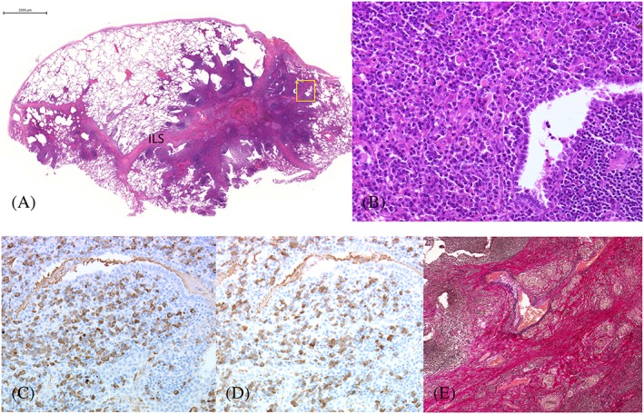
Figure 2.

Histopathology of the right S4. (A) Panoramic view of the right S4. Marked lymphoplasmacytic infiltration in the interlobular septa and alveolar walls with fibrosis and lymphoid follicles. (Hematoxylin‐eosin, ×1, bar 2 mm). (B) Prominent infiltration of plasma cells in the alveoli with scattered Russell bodies. (Hematoxylin‐eosin, ×20) (square on A). (C) κ‐positive plasma cells. (D) λ‐positive plasma cells. (×20) There was no monoclonality. (E) Granulomatous involvement of interlobular vein with disruption of elastic fibers. (Elastic van Gieson, ×10).
