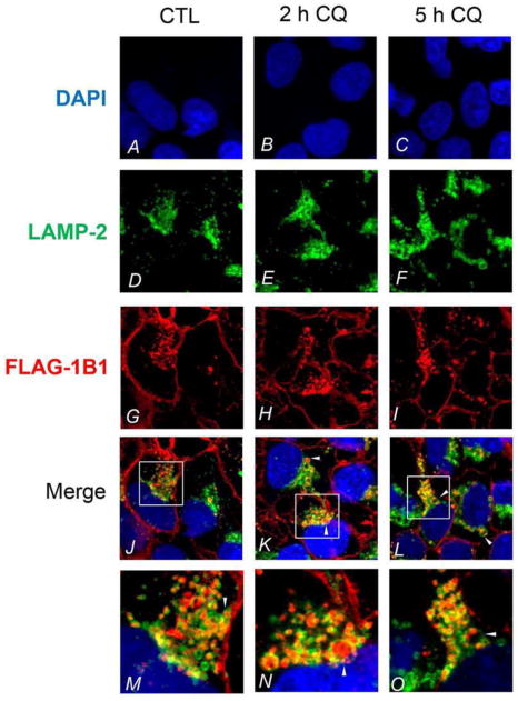
Figure 3.

Colocalization of FLAG-OATP1B1 and LAMP-2 in HEK293-FLAG-OATP1B1 cells. Coimmunofluorescence staining of FLAG-OATP1B1 (red) and LAMP-2 (green) was performed in HEK293-FLAG-OATP1B1 cells treated with vehicle CTL or 25 μM CQ for 2 and 5 h as described in the Experimental Section. White arrow heads indicate the accumulation of FLAG-OATP1B1 inside the LAMP-2-positive vacuoles. Nuclei were counterstained with DAPI (blue). Images were taken using Olympus FV1000 confocal microscope. Representative images from the same experiments are shown (3 and 2 separate experiments for 2 h and 5 h treatment, respectively).
