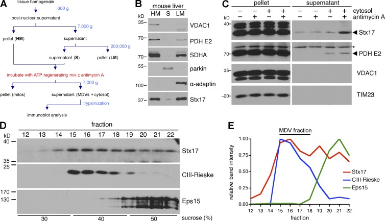
Figure 1.
Stx17 is biochemically enriched on mitochondrial-derived vesicles (MDVs). (A) Flowchart of cell-free MDV budding assay (see Materials and methods for complete details). (B) Mouse liver was fractionated (see Materials and methods), and 40 µg heavy-membrane (HM), soluble (S), and light-membrane (LM) fractions were separated by SDS-PAGE and analyzed by immunoblotting. (C) Trypsinized supernatants and pellets from in vitro MDV budding assays incubated in the presence of cytosol and/or 50 µM antimycin A (anti A) were separated, along with the reaction pellets (mitochondria), by SDS-PAGE and analyzed by immunoblotting. The asterisk indicates a nonspecific band. (D) A large-scale budding reaction (incubated with cytosol and 50 µM antimycin A) was fractionated over a discontinuous sucrose gradient, separated by SDS-PAGE and analyze by immunoblotting. (E) Intensity profiles for the proteins probed for in D, expressed as a fraction of their maximum intensity.

