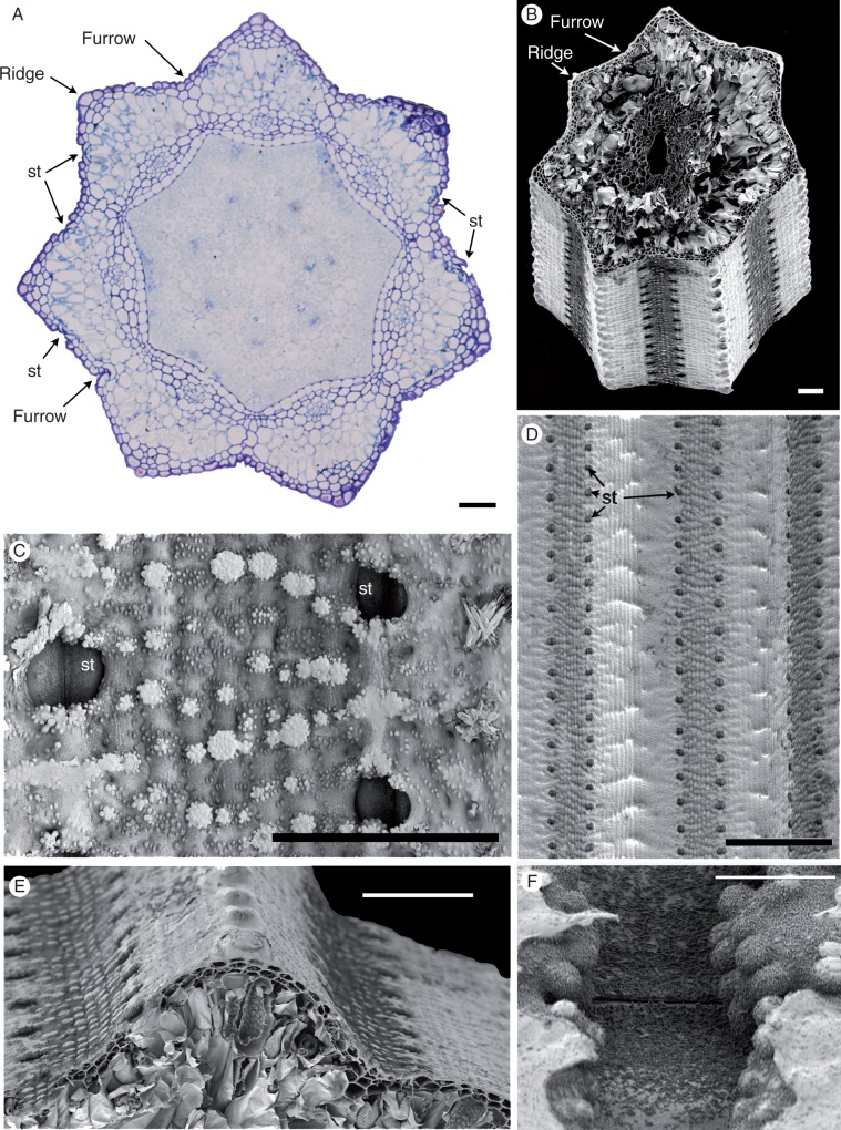
Fig. 2.
Equisetum myriochaetum (A, LM; B–F, SEM). (A) Transverse section of the stem just above a node, showing encircling fused leaf sheaths around the stem. (B). Transverse section of the stem at the internode. (C) Details of the stem surface with sunken stomata and silica. (D, E) Stem surface showing rows of stomata midway between ridges and furrows. (F) View of a sunken (closed) stomatal pore with fine epicuticular wax flakes. St, stomata. Scale bars = 100 μm in (A), (D), (E), 10 μm in (B), (F), 50 μm in (C).

