Abstract
Context:
Thermal insult is the major cause of thermal injury or death and in case of death due to thermal injury the body often has to be recovered from the site. Histologically, one can predict whether the victim was alive or dead when the fire was on going. However, determination of probable cause of thermal insult to which victim subjected to be difficult when the victim's body is found somewhere else from the crime scene or accident site or found alone. Hence, histopathological evaluation of the tissue which has undergone thermal insult in such conditions could help to place evidence in front of law officials, regarding probable condition, or scenario at time of burn of victim.
Aims:
Keeping this as a criteria in this study we aim to evaluate burnt tissue histopathologically, that undergone various degree of thermal insult, which simulates various real life scenario for mortality in burn cases.
Settings and Design:
We evaluate the changes in hematoxylin and eosin staining pattern of tissue which has undergone thermal insult compared to normal tissue and also the progressive changes in staining pattern, architectural, and cellular details.
Materials and Methods:
Samples were taken from the patients, in various surgical procedures. Each sample was cut into five parts with close margins so that each burnt tissue is evaluated for same field or region. The tissue that obtained was immediately subjected to varying degree of temperature over a specific period so as to simulate the various real-life condition. Then the tissues were fixed, processed, and stained with routine H and E staining. The processed slides of tissue were examined under the microscope, and the staining, and architectural changes were evaluated and described.
Results:
Results show that there was a progressive changes in the architectural pattern of the epithelium and connective tissue showing cleft formation and vacuolization, staining pattern also shows mixing of stains progressively as the severity of thermal insult increases.
Keywords: Burnt tissue, forensic pathology, gunshot injury, hematoxylin and eosin
Introduction
Forensic science is simply defined as the application of the science of law or legal matter.[1,2] A forensic investigation requires a skillful blend of science using both proven technique and common sense. The ultimate effectiveness of scientific investigation depends upon the ability of the forensic scientist to apply the scientific method to reach a valid, reliable, and a supportable conclusion about a question of controversy. Forensic is derived from the Latin word forum.[1] Forensic means legal: A word that means “to the forum.” The forum was the basis of Roman law and was a place of public discussion and debate pertinent to the law.[3,4]
The general definition of forensic dentistry is that it is a combination of sciences and art of dentistry and legal system, a crossroad of dental sciences and law. Forensic dentistry or more professionally forensic odontology is now integral part of judicial system for well over three decades. Overall, forensic odontology includes multiple area of investigation for gathering and interpretation of dental and related evidence within the overall field of criminalistics. Forensic dental evidence ranges from identification of person using dental records to the identification and analysis of bite marks on objects such as food stuff or on body surface of victim, along with an estimation of age using dental evidence.
Described as a medical trunk that serves the administration of justice, forensic science has different branches. Forensic pathology is probably the most emblematic one.[5] Forensic pathology is a subspecialty of pathology that focuses on determining the cause of death by examining a corpse.[6]
In today's fast-paced society, no matter how careful one is there is the chance that someone will be injured in an accident that was caused by negligence or plan that might lead to death. The cause could be due to multiple reasons - physical, chemical thermal insult. The majority of fatalities arising from thermal burns occur in intentional cases of burns like suicide, homicide which could be dowry-related, or accidental like accidental such as house fires, car accidents fires, forest fire, airplane crash fire, and hot air balloon fire.
Thermal insult is the major cause of thermal injury or death and in case of death due to thermal injury the body often has to be recovered from the site. Evidence collection from the body is the domain of the medical examiner in most jurisdictions. One of which is - examination of tissue (autopsy). Histologically, one can predict whether victim was alive or dead when fire was on going. However, determination of probable cause of thermal insult to which victim subjected to be difficult when the victim's body is found somewhere else from the crime scene or accident site or found alone. Hence, histopathological evaluation of the tissue which has undergone thermal insult in such conditions could help to place evidence in front of law officials, regarding probable condition, or scenario at time of burn of victim. Keeping this as a criteria in this study we aim to evaluate burnt tissue histopathologically, that undergone various degree of thermal insult, which simulates various real life scenario for mortality in burn cases. We evaluate the changes in hematoxylin and eosin (H and E) staining pattern of tissue which has undergone thermal insult compared to normal tissue and also the progressive changes in staining pattern, architectural, and cellular details in tissues subjected to increasing severity of thermal insult. We also correlate the type of injury as evaluated by H and E staining pattern with mode of injury.
Materials and Methods
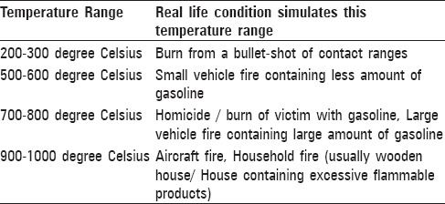
This is a prospective study and carried out in the Department of Oral Pathology and Microbiology at Sharad Pawar Dental College, DMIMS, Sawangi, and Wardha. Samples were taken from the patients, who came to our college for extraction gingivectomy, crown lengthening procedure, etc., Each sample was cut into five parts with close margins so that each burnt tissue is evaluated for same field or region. The tissue that obtained was immediately subjected to varying degree of temperature over a specific period so as to simulate the various real-life conditions [Table 1]. Then the tissues were fixed, processed, and stained with routine H and E staining. The processed slides of tissue were examined under the microscope, and the staining, and architectural changes were evaluated and described.
Table 1.
Various real life conditions/scenario and their temperature ranges
Results
Thirty samples, which were sectioned into five parts and blindly evaluated under light microscope, following results were obtained [Table 2 and Figures 1–16].
Table 2.
Histopathological changes in tissue under different temperature ranges

Figure 1.
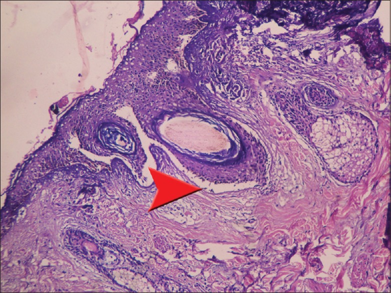
Cleft formation at junction of epithelium and connective tissue (arrowhead)
Figure 16.
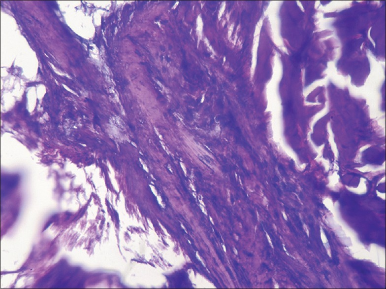
Mixing or running of stains
Figure 2.
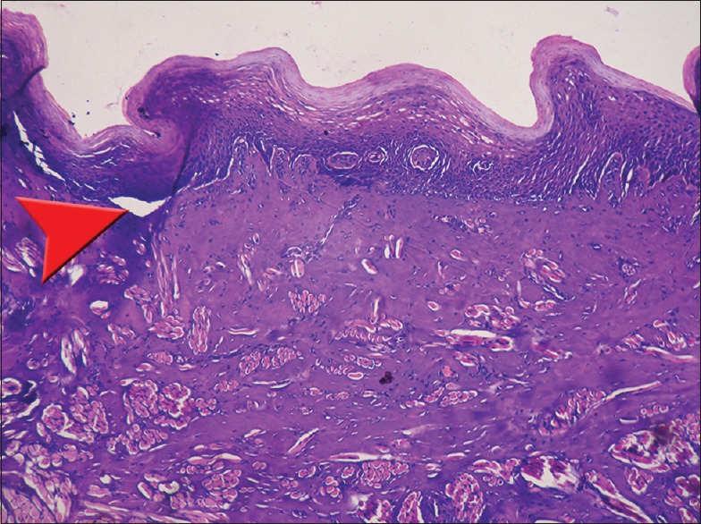
Epithelium shrink collagen begins to hyalinized (arrowhead)
Figure 6.
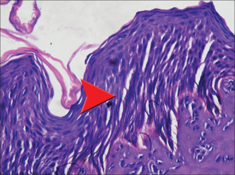
Separation extends up to basal, suprabasal layer and granular cell layer (arrowhead)
Figure 10.
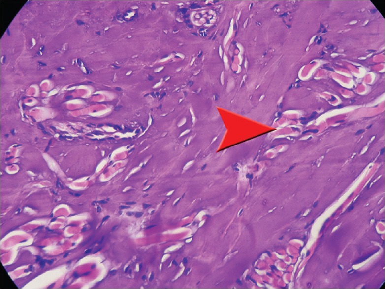
Homogenization of connective tissue and smudging of collagen fibres (arrowhead)
Figure 14.
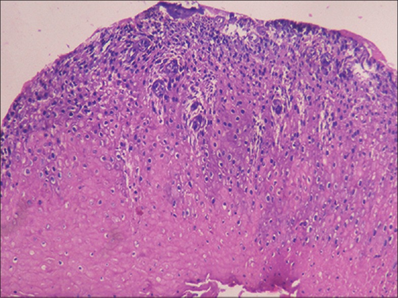
Normal H and E staining pattern with slight decrease in intensity of basophillia
Discussion
Thermal burns and related injuries are major cause of death and disability. Even in developed countries more than two million individuals annually are burned seriously and require medical treatment.[7,8] Fire deaths are some of the most challenging fatalities, both from the investigative and the autopsy aspect. The magnitude of deaths due to burns is as large as India is the only country in the world where fire is classified among the fifteen leading causes of death in 1998 standing 14th in the list.[7,9] Burn deaths are an important public health and social problem in India. There should be urgent need to implement burn prevention program in India which should aim at attending the incidence of burn injuries and mortality among young generation, especially in females. Burn has been reported to be the second most common cause of death in all medico-legal cases.[7] Burn injury is a complex process due to additive effects of inadequate tissue perfusion, free radical damage, systemic alterations in the cytokine milieu leading to protein denaturation and necrosis. The cellular and tissue changes are essentially those of hypoxic injury characterized by failure of multiple organ system.[7]
Victim remains at fatal fire scenes are typically difficult to detect, recover, and handle. All of the burned material at the scene, including biological tissue, is often modified to a similar appearance. Lack of on-scene recordation of relevant information related to body positioning and contextual relationships of remains as well as other physical evidence at the scene, further complicate trauma analysis, biological profile estimation, and event reconstruction. The present study addressed these problems by linking rigorous scene recovery and documentation methodologies with subsequent laboratory analyses of heat-altered human remains from fatal fire scenes.
On evaluating the histopathological changes in temperature range of 200–300°C that simulates a bullet shot of pistol from a contact range,[11] the following changes in H and E stained tissue section occurs such as cleft formation at junction of epithelium and connective tissue [Figure 1], initial separation between cells in basal and suprabasal layer of epithelium [Figure 5], homogenization of connective tissue with separation of connective tissue [Figure 9], normal H and E staining but slight decrease in intensity of basophillia [Figure 13]. The probable reason for such changes could be due to initial unequal shrinkage of tissue, dehydration of tissue leads to altered staining.
Figure 5.
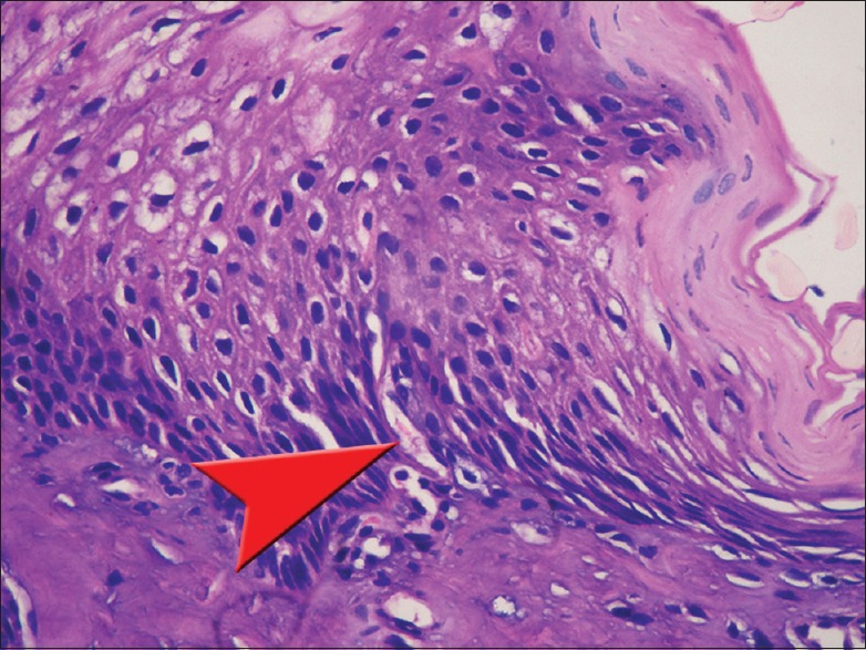
Initial separation between cells in basal and suprabasal layer (arrowhead)
Figure 9.
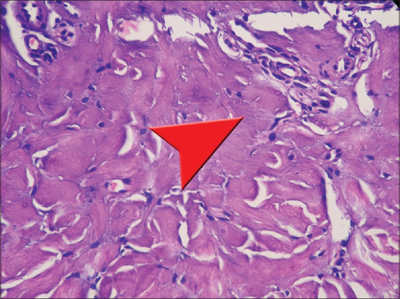
Homogenization of connective tissue with separation of connective tissue (arrowhead)
Figure 13.
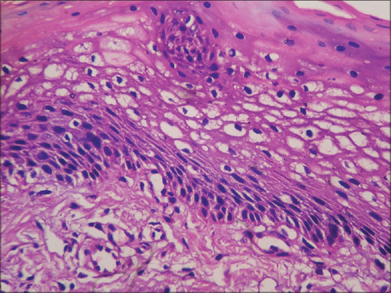
Normal H and E staining pattern with slight decrease in intensity of hematoxylin
On evaluating the histopathological changes in temperature range of 500–800°C that simulates real-life car accidents burns[12] or from a bullet shot a rifle,[11] the following changes in H and E stained tissue section occurs such as total epithelium separation [Figure 3], mildly obscured and disorganized cellular details [Figure 7], vacuolization of connective tissue [Figure 11], and decrease in eosinophilia [Figure 15], which could be due to unequal shrinkage of tissue, rupture of vacuole-bound structure, damage to the cell membrane cause leaking of cytoplasmic fluid from cell that probably alters the pH of tissue and finally leads to alteration in staining respectively.
Figure 3.
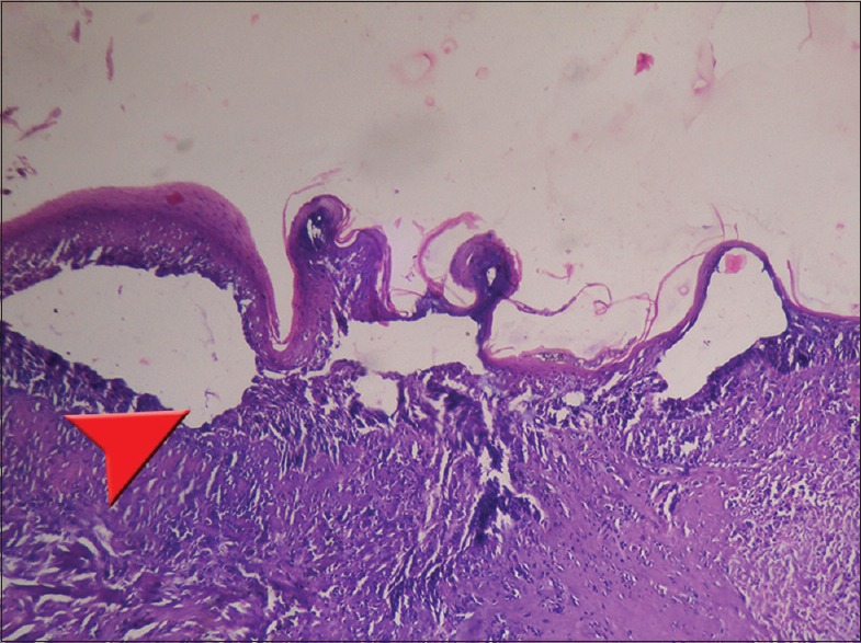
Epithelial separation from connective tissue (arrowhead)
Figure 7.
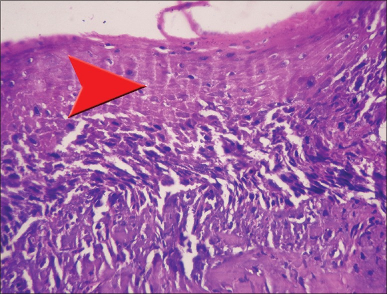
Cellular details are mildly obscured and disorganized (arrowhead)
Figure 11.
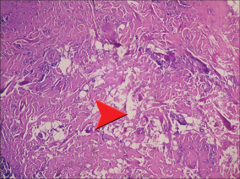
Vacuolization of connective tissue (arrowhead)
Figure 15.
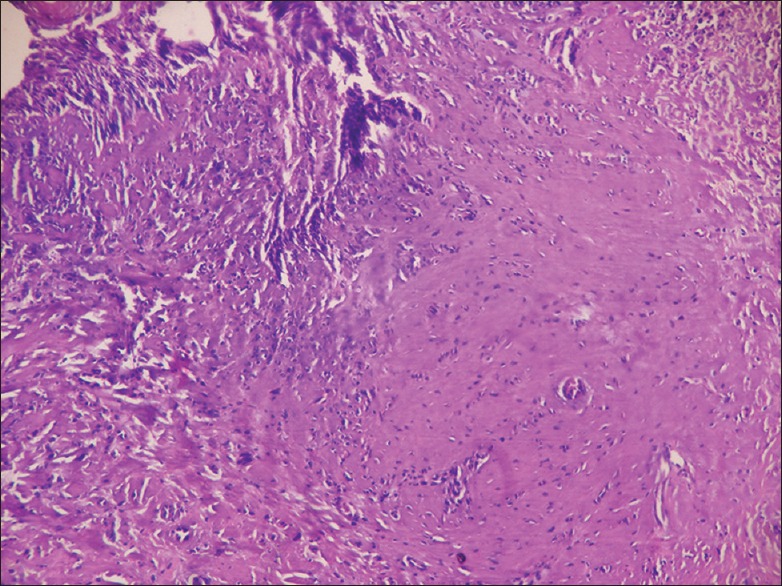
Decrease in intensity of eosin staining
On evaluating the histopathological changes in temperature range of 1000–1200°C, which simulates the conditions such as household fires [Figure 5],[13] gasoline fires,[12,13] plane crashes, heavy machine gun bullet.[11] The following changes in H and E stained tissue section occurs like total epithelial separation from connective tissue [Figure 4], totally obscured cellular detail [Figure 8], vacuolization of connective tissue [Figure 12] and mixing or running of stains [Figure 16]. The probable reason for these changes could be due to unequal shrinkage of tissue, damage to the cell membrane cause leaking of cytoplasmic fluid from cell. While, mixing of stains is probably due to leaking of nuclear material into cytoplasm of cell due to rupture of nuclear membrane.
Figure 4.
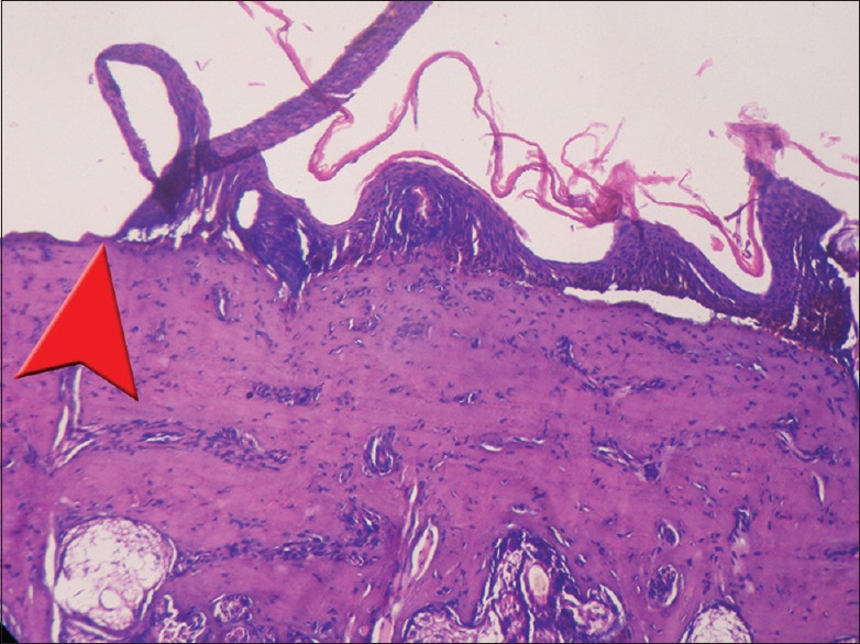
Total epithelial separation from connective tissue (arrowhead)
Figure 8.
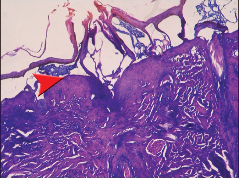
Cellular details are totally obscured (arrowhead)
Figure 12.
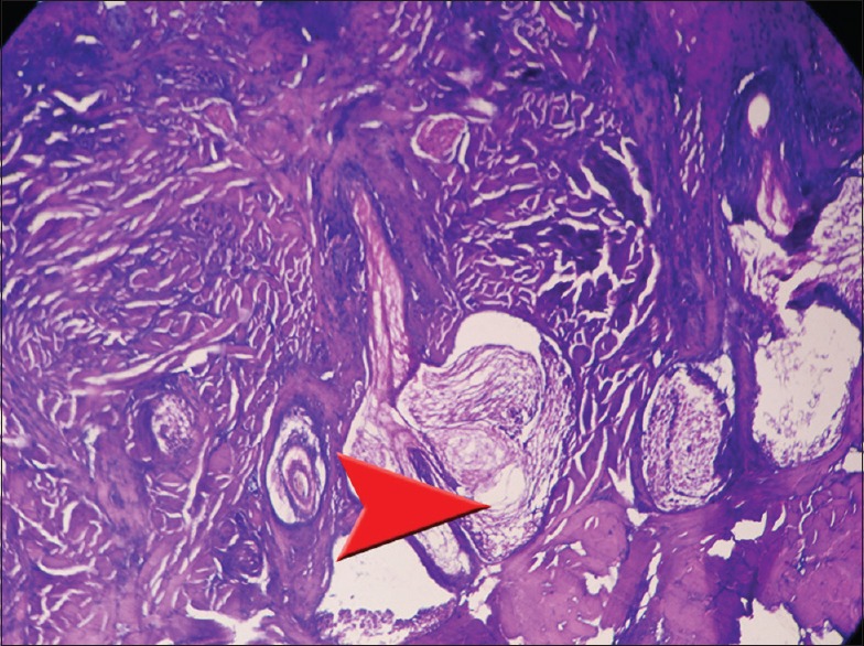
Vacuolization of connective tissue and destruction of salivary and sebaceous gland (arrowhead)
All the histopathological changes corresponding to different temperature ranges that simulates to different real life situations probably gives us an additional diagnostic aid to link the rigorous scene recovered tissue and its documentation with subsequent laboratory analyses from fatal fire scenes. As there is no such studies found which is been used to compare the results of this study, thus this study may provide an astringent pathway to rule out the possible cause of death by burns and to reach to the proper justice.
Conclusion
Human body can be injured by various thermal sources at several temperature ranges. Most commonly the human body suffers thermal insult in household fire, car, or plane accident and even by homicide or suicide. One can predict the possible scenario of the fire or burn when the victim's burnt body is found at the site of accident or scene. However, the issue becomes more complex if the victim is found far away from the site of the scene. In such condition, it becomes essential to ascertain the exact cause of death and correlate the extent of tissue damage with the proposed cause of death. Therefore, this study is an attempt to tabulate and correlate the severity of thermal insult, and the tissue damage observed. In cases where there is destruction of evidence or deliberate misleading of investigating personnel by concealment of actual site and mode of injury. Histopathological analysis of tissue can play a valuable part in helping the forensic pathologist to arrive at a correct conclusion.
Financial support and sponsorship
Nil.
Conflicts of interest
There are no conflicts of interest.
References
- 1.Gupta M, Pachauri A, Mishra P, Singh P, Mishra R. New horizons for forensic dentist. Indian J Compr Dent Care. 2014;4:1. [Google Scholar]
- 2.Funayama M, Kanetake J, Ohara H, Nakayama Y, Aoki Y, Suzuki T, et al. Dental identification using digital images via computer network. Am J Forensic Med Pathol. 2000;21:178–83. doi: 10.1097/00000433-200006000-00017. [DOI] [PubMed] [Google Scholar]
- 3.Gupta S, Agnihotri A, Chandra A, Gupta OP. Contemporary practice in forensic odontology. J Oral Maxillofac Pathol. 2014;18:244–50. doi: 10.4103/0973-029X.140767. [DOI] [PMC free article] [PubMed] [Google Scholar]
- 4.KeiserNeilsen S. Person Identification by Means of Teeth. 1st ed. Bristol: John Wright and Sons; 1980. p. 190225. [Google Scholar]
- 5.Pinheiro J. Introduction to forensic medicine and pathology from forensic anthropology and medicine. Totowa, NJ: Copyright Humana press; 2006. [Google Scholar]
- 6.Williams DJ, Ansford AJ, Priday DS, Forrest AS. Forensic Pathology, Colour Guide. Edinburgh: Churchill Livingstone; 1998. pp. 1–7. [Google Scholar]
- 7.Shinde AB, Keoliya AN. Study of burn deaths with special reference to histopathology in India. Indian J Basic Appl Med Res. 2013;2:1153–9. [Google Scholar]
- 8.Cakir B, Yegen BC. Systemic response to burn injury. Turk J Med Sci. 2004;34:215–6. [Google Scholar]
- 9.Batra AK. Burn mortality: Recent trends and sociocultural determinants in rural India. Burns. 2003;29:270–5. doi: 10.1016/s0305-4179(02)00306-6. [DOI] [PubMed] [Google Scholar]
- 10.Symes SA, Dirkmaat DC, Ousley S, Chapman E, Cabo L. Recovery and Interpretation of Burned Human Remains, Document No: 237966 [Google Scholar]
- 11.Mark A. Finney, Trevor B. Maynard, Sara S. McAllister, Ian J. Finney. A Study of Ignition by Rifle Bullets; Publications Distribution Rocky Mountain Research Station 240 West Prospect Road Fort Collins, CO 80526 Rocky Mountain Research Station, Research Paper RMRS-RP-104, August 2013 [Google Scholar]
- 12.Motor Vehicle Fires; Walters Forensic Engineering. Toronto, ON: [Last accessed date 2014 Nov 18]. Available from: http://engineering@waltersforensic.com . [Google Scholar]
- 13.Kerber S. Analysis of Changing Residential Fire Dynamics and Its Implications on Firefighter Operational Time Frames [Google Scholar]


