Figure 1.

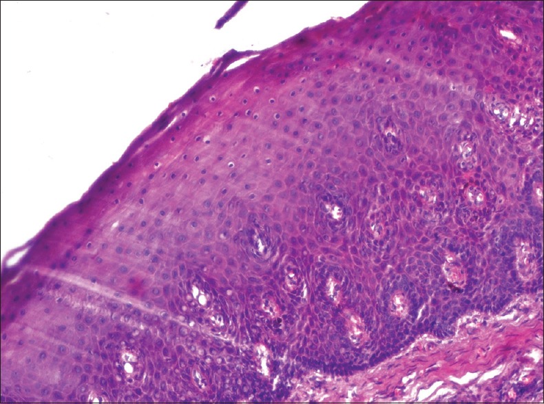
Photomicrograph showing histological changes in an antemortem unfixed gingival tissue at 15 min (H and E, ×200) isolated cells showing chromatin clumping, nuclear vacuolation, and prominent and widened intercellular bridges

Photomicrograph showing histological changes in an antemortem unfixed gingival tissue at 15 min (H and E, ×200) isolated cells showing chromatin clumping, nuclear vacuolation, and prominent and widened intercellular bridges