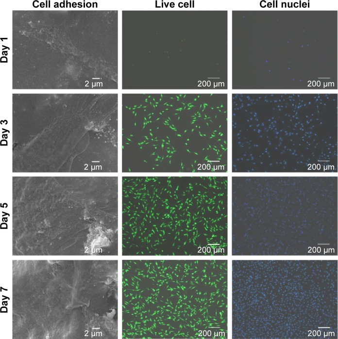
Figure 8.
SEM images and fluorescence microscopy images (counterstained with calcein-AM and DAPI for live cell and cell nuclei in green and blue, respectively) of MG-63 cells on S5 scaffold after 1 day, 3 days, 5 days, and 7 days of culture.
Notes: Live cells appeared as bright green dots and related cytoblast as purplish blue dots. S5, PEEK–10 wt% nano-HAP–0.2 wt% GNSs–0.8 wt% CNTs. Abbreviations: DAPI, 4,6-diamidino-2-phenylindole; SEM, scanning electron microscopy; CNTs, carbon nanotubes; GNSs, graphene nanosheets; HAP, hydroxyapatite; PEEK, polyetheretherketone.

