Figure 1.
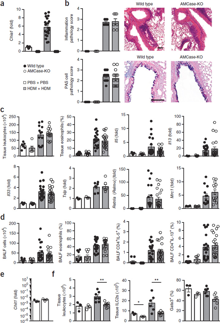
AMCase-deficient mice develop acute HDM-induced lung allergy despite diminished ILC2s. (a) Quantitative PCR analysis of AMCase (Chia1) gene expression in lung tissue from wild-type or AMCase-deficient mice (AMCase-KO) sensitized and challenged intranasally with PBS (n = 9 mice per genotype) or house dust mite (HDM) (n = 21 mice per genotype), expressed as fold change relative to expression of PBS-treated wild-type mice. (b) Lung pathology of mice in a. At left, scores (scale 0–4, with 4 being the worst) of tissue sections stained with hematoxylin and eosin (inflammation; PBS, n = 3; HDM, n = 7) and periodic acid-Schiff (PAS; PBS, n = 6; HDM, n = 15; pooled from two experiments). At right, representative staining (scale bars, 100 µm). (c) Lung tissue leukocyte (PBS: n = 6, HDM: n = 15 pooled from two experiments) and eosinophil quantification (PBS: n = 9, HDM: n = 22; pooled from three experiments) from three lung lobes of mice in a and gene expression analysis (PBS: n = 9, HDM: n = 22; pooled from three experiments). (d) Leukocyte and eosinophil quantification and intracellular cytokine analysis of lymphocytes collected from bronchoalveolar lavage fluid (BALF) of mice in a (PBS, n = 9; HDM, n = 22). (e) Chitotriosidase gene expression in lung tissue (representative of naive and allergic lungs) (wild type, n = 3; AMCase-KO, n = 3) shown relative to expression of the housekeeping gene Rplp2. (f) Following only sensitization of wild-type or AMCase-KO mice with intranasal PBS (n = 3) or HDM (n = 7), quantification of total lung leukocytes, total lung IL-4+IL-13+ ILC2s, and Gata3+ cells among ILCs in the lung. Data are pooled from three experiments (a,d) or are from one experiment representative of three independent experiments with similar results (e,f). Error bars, s.e.m.; each data point represents a value for an individual mouse.

