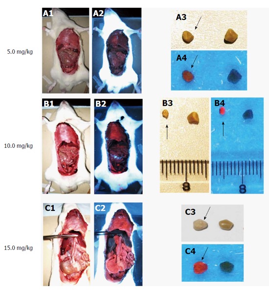
Figure 7.

Macroscopic digital imaging under white and UV (365 nm) lights in rats of cholelithiasis receiving intravenous 1 × 10⁻³ M hypericin at 5, 10, 15 mg/kg and their corresponding extracted gallstones. Rats receiving hypericin at 5 mg/kg (A1) exhibited almost no fluorescence in organs (A2). The extracted gallstones (black arrow) showed faint fluorescent relative to control stones (A3, A4). With 10 mg/kg hypericin (B1), moderate fluorescence appeared in the diaphragm (B2). Higher red fluorescence in gallstones (black arrow) was observed as compared to control stone (B3, B4). Rats treated with 15 mg/kg hypericin (C1) revealed high fluorescence in visceral organs (C2). The stone (black arrow) displayed high fluorescence as compared to the control (C3, C4).
