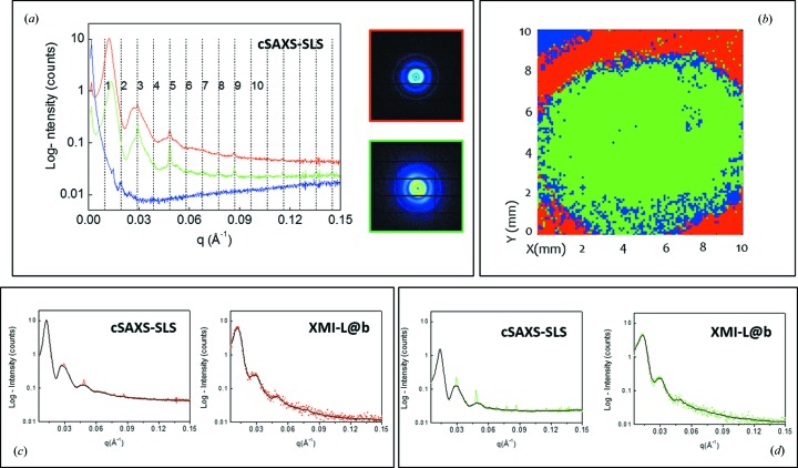
Figure 1.
(a) cSAXS-SLS reference data, as selected by adaptive binning from the cSAXS-SLS data set: blue background, red and green SAXS profiles corresponding to the two-dimensional SAXS data of the insets; (b) results of the canonical correlation analysis on the explored area (cSAXS-SLS data set), with the red color corresponding to the red profile in (a) and marking the outer corona without epithelium layer and the green color corresponding to the green profile in (a) in the center of the cornea; (c) cSAXS-SLS (left) and XMI-L@b (right) reference data for the red profile marking the outer corona without epithelium layer; (d) cSAXS-SLS (left) and XMI-L@b (right) reference data for the green profile marking the center of the cornea. Black lines are the filtered cSAXS-SLS and de-noised XMI-L@b reference data.

