Figure 1.
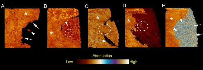
A = Empty negative control defect showing a minimum of healing with uneven, centric ingrowth from the margins of the defect (arrows) and small bone islets in the center of the defect (arrowhead). Mature intact calvarial bone marked with asterisk.
B = Defect filled with combination of particulated autogenous bone (yellow and orange granules inside of dotted circle) and fibrin glue (dark red areas pointed with arrowhead). Mature intact calvarial bone marked with asterisk.
C = Defect with autologous bone block. The margins of the bone block have been partially resorbed (long arrow) and partial osseous continuity of the margin the bone block can be seen (inside of dotted circle). Mature intact calvarial bone marked with asterisk.
D = Resorbable BAG scaffold in defect (dark red). Ingrowth of new bone from the defect margin can be seen as well as very small ossifying spots in the middle of scaffold material (yellow/orange spots inside of dotted circle). Mature intact calvarial bone marked with asterisk.
E = TCP granules (blue spots pointed with arrowheads) filling a calvarial defect. Bone formation on the material surfaces can be seen (yellow and orange spots pointed with arrows). Mature intact calvarial bone marked with asterisk.

