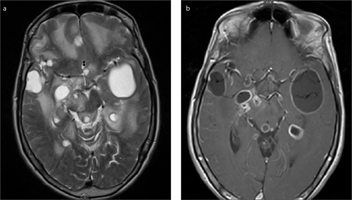
Figure 1. a, b.
T2-weighted image shows multiple cystic lesions with oedema in the subarachnoid space and subpial area (a). Postcontrast T1-weighted image shows rim-like contrast enhancement on the lesion walls, however, mural nodule contrasts are also observed on cyst walls in some cystic lesions (b).

