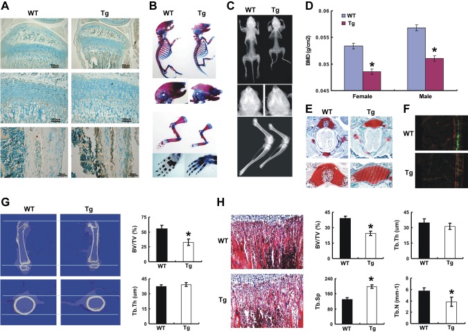
Figure 4.
Targeted overexpression of ECM1 in osteoblast leads to short-limbed dwarfism and a delay in endochondral ossification in young mice. A) Increased ECM1 expression in newborn transgenic mouse osteoblasts detected by immunohistochemistry. B) Skeletal preparation with alcian blue/alizarin red staining of newborn WT and TG mice. Representative pictures shown as whole-mount view and high magnification of forelimbs and hind limbs. C) Representative pictures of whole body from WT and ECM1 transgenic mice at 3 wk old assayed by X-ray. D) Dual-energy X-ray absorptiometry scan analysis for the total bone mineral density (BMD) in WT and transgenic mice at 3 wk; n = 6 in each group. E) Delayed formation of ossification center in transgenic mice as detected by safranin O staining of the vertebra of 3-wk-old mice. F) Calcein double labeling in 4-wk-old mice. Bone formation rate (BFR) represented by the distance between 2 green labels, and BFR was compared between WT and transgenic mice; n = 5 for each group. G) Coronal section of middle femurs were collected and bone quality was compared in 3-wk-old WT and transgenic mice assayed by micro-CT. H) Morphologic assessment of bone quality was performed in the trabecular bone of tibia from WT and ECM1 transgenic mice, and parameters were statistically analyzed. Tb.N, trabecular number; Tb.Sp, trabecular separation; Tb.Th, trabecular thickness. Data show means ± sem. *P < 0.05 between WT and transgenic mice; n = 6. Original magnification, ×40.

