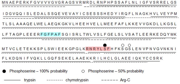
Figure 5.
Sequence coverage of phosphorylated hBVR by proteolysis and mass spectrometry. Products of kinase reaction that included Akt1 and his6-hBVR (purified from E. coli) were resolved by gel electrophoresis. Three identical samples, each containing 2 µg of protein, were digested with trypsin, chymotrypsin, or Arg-C protease. Products were identified by mass spectrometry. Extent of sequence coverage by each digest is indicated by underlining; serine residues identified as phosphorylation sites are indicated by dots above sequence. Red shading indicates Akt binding/phosphorylation site as predicted by computational analysis, and blue shading indicates RFxFPxFS PDK1 binding motif.

