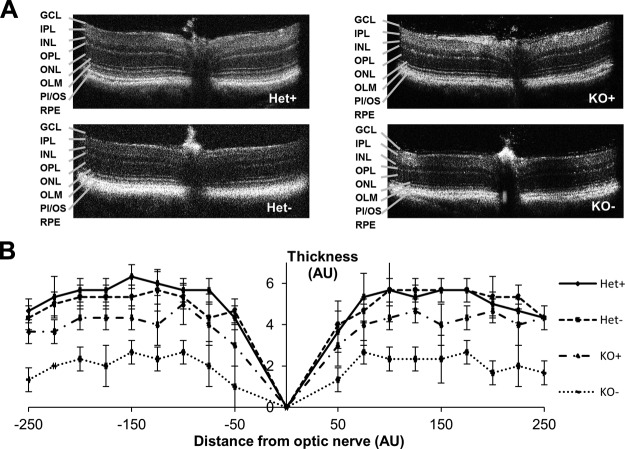
Figure 4.
Dietary vitamin A deprivation in KO mice results in thinning of the photoreceptor layer compared with Het mice raised on a VAS or VAD diet, or KO mice raised on a VAS diet. A) Typical OCT images of Het+, Het−, KO+, and KO− mice. B) Measurement of photoreceptor thickness from OCT images. AU, arbitrary units; GCL, ganglion cell layer; INL, inner nuclear layer; IPL, inner plexiform layer; OLM, outer limiting membrane; ONL, outer nuclear layer; OPL, outer plexiform layer; PI/OS, photoreceptor inner/outer segments. Data present values from n = 6 per genotype and dietary condition.

