Fig. 3.
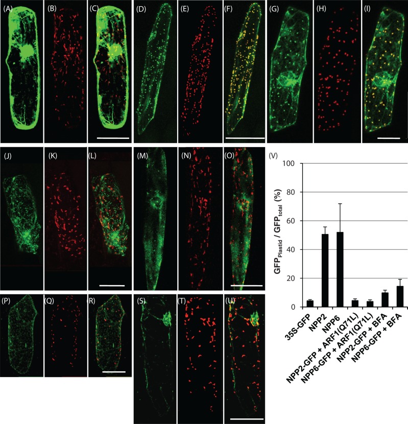
Expression and localization of NPP2–GFP and NPP6–GFP in onion epidermal cells. (A–C) Control experiments. Double labeling with GFP and WxTP–DsRed was performed in onion cells. (D–F) Onion cells expressing NPP2–GFP and WxTP–DsRed. (G–I) Onion cells expressing NPP6–GFP and WxTP–DsRed. (J–O) Effect of Arabidopsis ARF1(Q71L) on the localization of NPP2–GFP (J–L) and NPP6–GFP (M–O). (P–U) Effect of 90μM BFA on the localization of NPP2–GFP (P–R) and NPP6–GFP (S–U). (A), (D), (G), (J), (M), (P) and (S) GFP, green; (B), (E), (H), (K), (N), (Q) and (T) WxTP–DsRed, red; (C), (F), (I), (L), (O), (R) and (U) GFP and DsRed merged. (A–U) Projections of stacks of 20–30 images per cell, acquired from the top to the middle of the cell, every 1.5μm. Scale bars = 100μm. (V) Statistical evaluation of the plastidial localization of NPP2–GFP and NPP6–GFP. Ratios of the fluorescence intensity of GFP in the plastidial area to GFP in the whole cell (GFPplastid/GFPtotal) (%) were determined. Values are represented as mean ± SD (n = 4). NPP2–GFP and NPP6–GFP were evidently localized in the plastids visualized with WxTP–DsRed, and the plastid targeting of NPP2–GFP and NPP6–GFP was significantly prevented in cells expressing ARF1 mutants or BFA treatments.

