
Figure 2. Tau and amyloid PET imaging in AD.
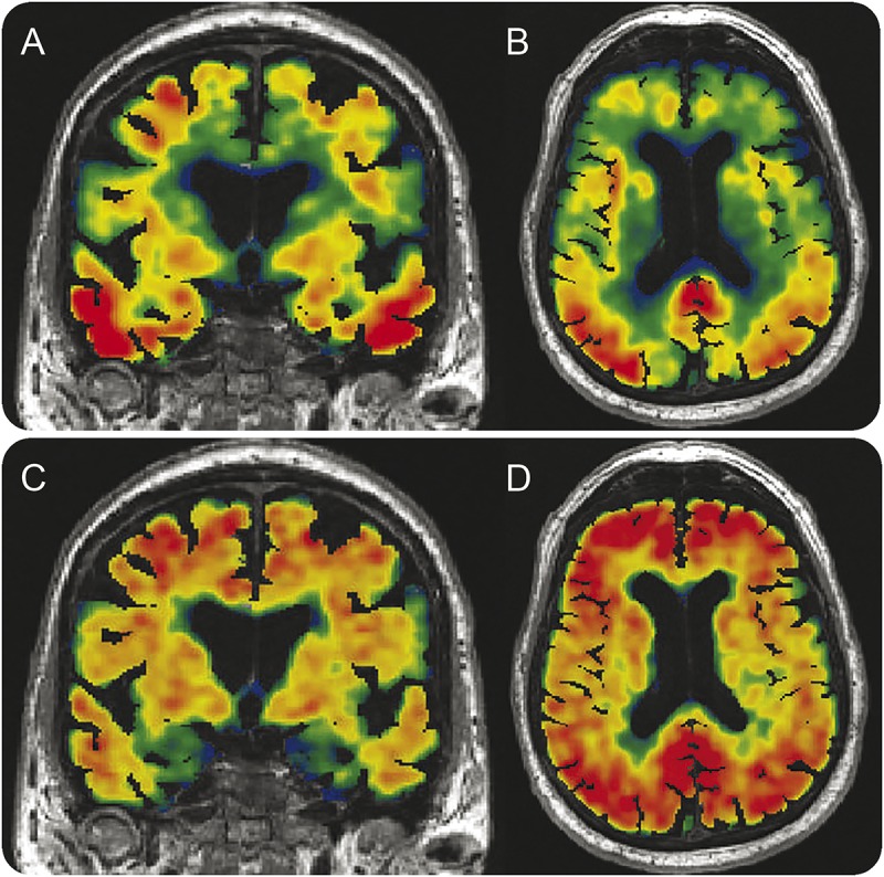
A 79-year-old man with a clinical diagnosis of AD dementia. He is a participant in the Mayo Alzheimer's Disease Research Center study. (A, B) Coronal and axial tau PET images (AV1451) superimposed on MRI. (C, D) Coronal and axial Pittsburgh compound B PET images superimposed on MRI. The tau PET images (top) illustrate extensive tracer uptake in basal lateral temporal, parietal, and frontal isocortex with sparing of sensory motor and primary visual cortices. Off-target binding is seen in the basal ganglia, which is characteristic of this tracer. Although areas of spatial overlap between the tau and amyloid tracers are present, abundant amyloid tracer uptake is seen in the frontal lobes, but not with the tau tracer. Conversely, abundant uptake is seen in the medial temporal lobes with the tau ligand but not with the amyloid ligand. AD = Alzheimer disease.