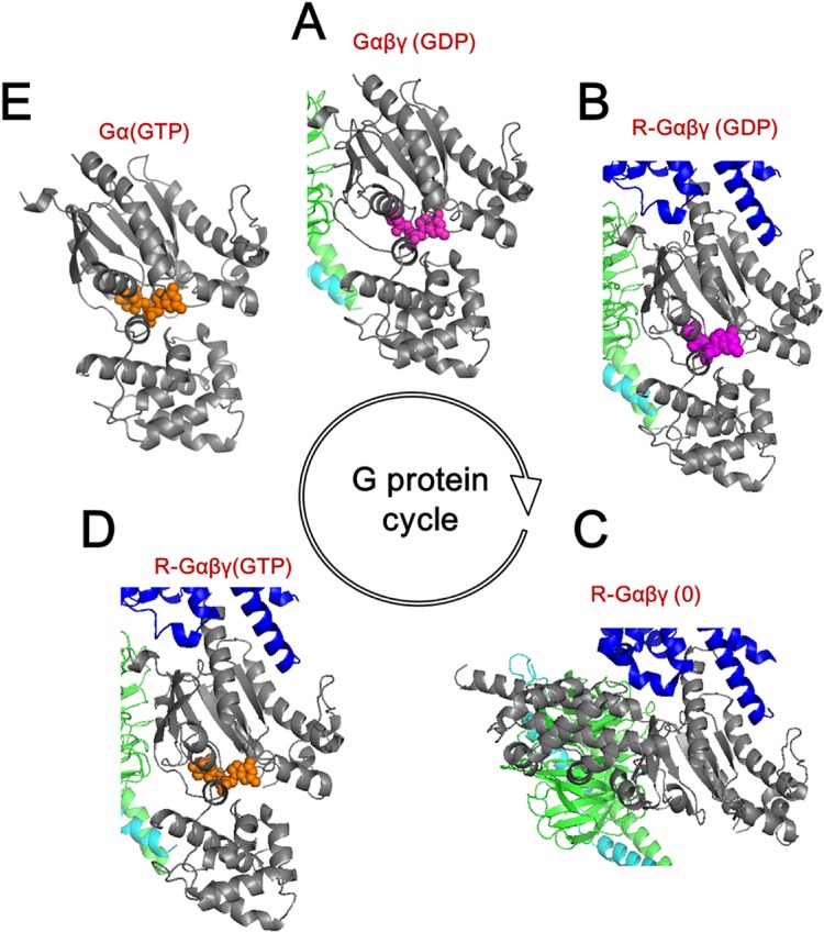
Fig 2. Overview of the G protein cycle.
(A) Inactive GDP (purple)-bound Gs protein heterotrimer with α (grey), β (green), and γ (cyan) subunits. (B) Activated receptor (blue)-bound temporal state of Gαβγ(GDP). (C) Receptor- bound fully opened Gαβγ. (D) The receptor- bound temporal state of Gαβγ with GTP (orange). (E) Active GTP-bound Gα. Receptor and Gβγ subunits were truncated for convenience.

