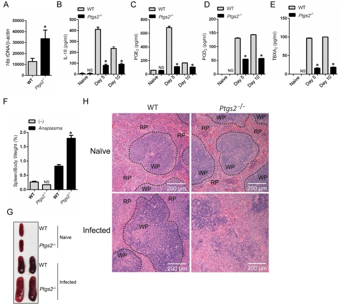
Fig 10. COX2 restricts A. phagocytophilum infection in vivo.
A. phagocytophilum infection of WT (n = 20) and COX2 (ptgs2) -/- (n = 10) mice. Bacterial load in the (A) peripheral blood of infected mice at day 15. (B) IL-18, (C) PGE2, (D) PGD2 and (E) TBXA2 release in the serum of infected animals. (F-G) Splenomegaly for COX2 (ptgs2 -/-) mice infected with A. phagocytophilum. (H) Splenic architecture depicting the red (RP) and the white (WP) pulp during A. phagocytophilum infection. One-way ANOVA-Tukey; Student t test; *P < 0.05. NS–not significant. (-) non-stimulated.

