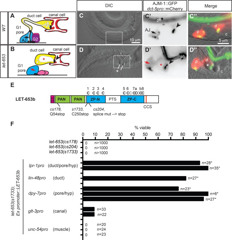
Fig 1. let-653 acts in the excretory duct and pore cells, not the canal cell.
(A-D) let-653 mutants have morphological defects in the duct and pore. (A,B) Schematics of excretory system morphology in WT (A) and let-653 mutants (B) at the early L1 stage. G1 pore cell is shown in blue, duct cell in yellow, canal cell in red, and G2 and W epidermal cells in pink. White regions represent lumens. Heavy black lines represent apical junctions. Black arrow, pore cell autocellular junction (AJ); red arrow, missing AJ in let-653 mutant. Arrowheads, duct-canal intercellular junction (IJ), *, duct lumen dilation. Anterior is to the left and ventral is down in all images. (C, D) Duct (d) and pore (p) morphology in WT and let-653(cs178) mutants. Cell bodies (red in C”, D”) and all apical junctions (green in C”, D”) are marked as labeled. DIC, differential interference contrast of head; box shows region magnified in subsequent panels. c, canal cell nucleus. ph, pharynx. (E) LET-653b protein schematic and locations of mutant allele lesions. SP, signal peptide. PAN, PAN-Apple domains. ZP, zona pellucida domain. Conserved ZP domain cysteines are indicated above in black; additional cysteines are indicted in grey. PTS, Proline/Threonine/Serine-rich region is predicted by Net-O-Glyc [99] to be highly O-glycosylated. LET-653b is the shortest of three protein isoforms produced from the let-653 locus by alternative splicing; the other isoforms contain a larger central PTS domain (www.wormbase.org). CCS, consensus furin cleavage site (R-X-R/K-R) [100]. (F) Tissue-specific expression of let-653b cDNA in the duct and/or pore efficiently rescued let-653(s1733) lethality. Data from multiple independent transgenic lines per construct are shown. *, p<0.01, Fisher’s Exact test, compared to non-transgenic siblings.

