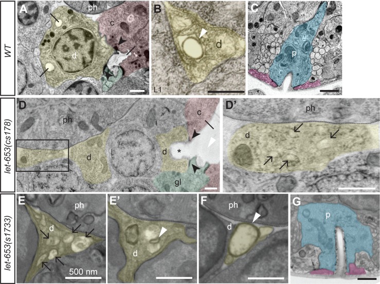
Fig 3. TEM analysis of let-653 mutant excretory system.
TEM sections of the excretory duct or pore region in WT (A-C) or let-653 mutants (D-G), pseudocolored to indicate the duct (d, yellow), G1 pore (p, blue), G2 (pink), canal cell (c, red) and excretory gland cell (gl, green). Dotted lines indicate basement membrane of the pharynx (ph). (A) Lateral section from a normal, late 3-fold stage embryo, showing the duct cell body and the luminal connection between the duct and canal cells. Black arrowheads indicate intercellular junctions, and lines indicate lumen. Multiple luminal segments appear in a single cross-section due to the winding path of the wild-type duct lumen. (B) Transverse section through the duct process in a normal L1 larva. White arrowhead indicates the cuticle lining of the duct lumen. (C) Excretory pore opening from the same animal as in A. (D, D’) Skewed lateral section through a let-653(cs178) late 3-fold stage embryo. A portion of the duct lumen (asterisk) and the canal cell lumen are greatly dilated behind a region devoid of lumen. Most of the duct cell body and the duct nucleus are outside the plane of this section; the central nucleus belongs to a different cell. Boxed region in D is magnified in D’; this region corresponds to the beginning of the duct process (compare to B). This region lacks a continuous lumen, but instead contains multiple smaller membrane-bound structures (black arrows). (E-F) Transverse sections from the duct processes of two let-653(s1733) L1 larvae. The lumen is fragmented (E) or contains abnormally small (E’) or large (F) rings of cuticle-like material (compare to B). (G) The G1 pore is intact and has a normal lumen diameter (compare to C) in this mutant L1. Scale bars, 500 nm.

