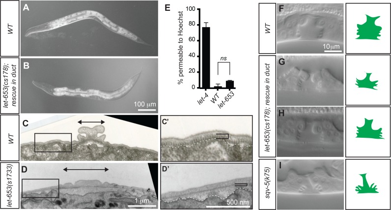
Fig 4. let-653 is required for proper alae formation and vulva lumen shaping.
(A,B) Comparison of body shape in WT and let-653(cs178) L4 larvae rescued to viability with a duct-specific lin-48pro::LET-653::SfGFP transgene. The mutants are slightly shorter and fatter than WT. (C,D) TEM images of alae in WT and let-653(s1733) L1 larvae. Transverse sections through the head region at the posterior bulb of the pharynx are shown. Boxed regions are magnified in adjacent panels. Arrowed bars indicate width of the alae. (C’, D’) let-653 mutants have alae and cuticle organization defects. White dotted line indicates edge of the epidermal cell layer. In let-653 mutants, the striated layer of the cuticle (black bracket) appears thin or absent, and the space between this layer and the underlying epidermis is expanded and filled with diffuse, lightly staining material. (E) The permeability barrier is intact in let-653 mutants. L1 larvae were incubated in 2 ug/ml Hoechst dye in M9 for 15 minutes and then rinsed and scored for nuclear fluorescence. let-4(mn105) was used as a positive control for permeability [38]. Data from three replicates shown, n>15 per genotype for each experiment. No significant difference found between let-653 and WT permeability by Student’s t-test, two-tailed. (F-I) Comparison of L4 vulva lumen morphology in WT (F), duct-rescued let-653 mutants (G,H) and a hypomorphic chondroitin biosynthesis mutant sqv-5(k175) [101]. Animals were staged as mid-L4 based on fusion of the anchor cell and horizontal orientation of the ventral vulva “fingers”. Modest vulval expansion defects or asymmetries were observed in 11/31 of mutant larvae.

