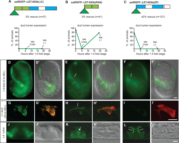
Fig 8. LET-653 PAN and ZP domains confer distinct localization patterns and mediate duct lumen cycling and protection, respectively.
(A-C) LET-653 fusions lacking the C-terminal, ZP or PAN domains showed differential functions and localization in the duct. Only LET-653(ZP) rescued mutant defects. (D,G,J) LET-653(∆C) accumulated intracellularly rather than extracellularly, suggesting a failure in secretion. (E-F) LET-653(PAN) and LET-653(ZP) fusions localized normally to the duct in 1.5 fold embryos. (H-I) LET-653(PAN) accumulated in the late L1 larval duct lumen, but LET-653(ZP) did not. Confocal slices. (G’,H’,I,) lin-48pro::mRFP marks the duct cell. (K) LET-653(PAN) associated with fibrous material in the center of the lumen (arrow) (L) LET-653(ZP) associated with the dorsal apical membrane region. D’,E’,J’,K’,L’ show DIC images for comparison. Scale bars, 5 μm.

