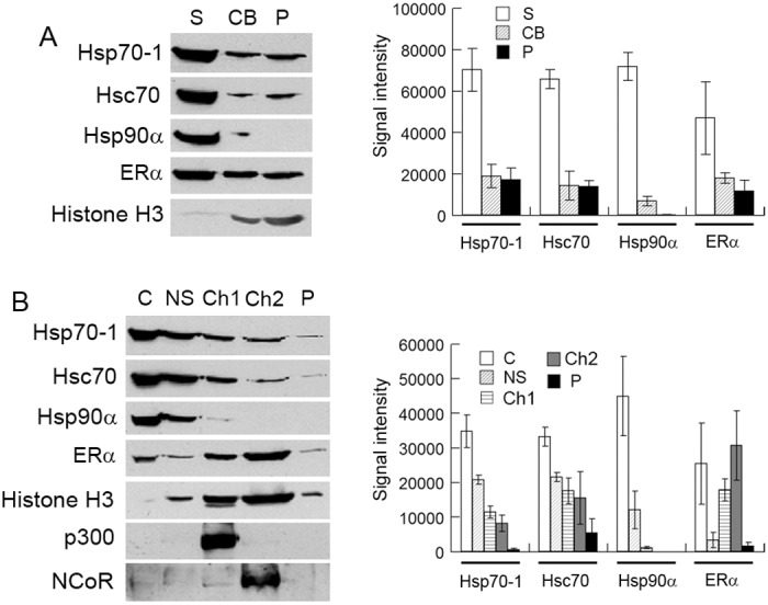
Fig 3. Hsp70-1 and Hsc70 are associated with chromatin.

(A) The MCF7 cell extract was fractionated into soluble protein (S), chromatin-binding protein (CB), and the pellet (P), and then analyzed by Western blotting with the indicated antibodies (left panel). Right panel, quantification of Western blots. (B) The MCF7 cell extract was fractionated into cytoplasmic protein (C), nuclear soluble protein (NS), transcriptionally active chromatin (Ch1) and inactive chromatin (Ch2), and analyzed by Western blotting with the indicated antibodies (left panel). Right panel, quantification of Western blots. Histone H3, p300, and NCoR were used as markers of chromatin-binding protein, active chromatin, and inactive chromatins, respectively. Signal intensity values in the Western blot quantifications were arbitrary numbers obtained by analyzing the protein bands with ImageJ software. Values in the Western blot quantifications were the means ± S.D. of three separate sample preparations.
