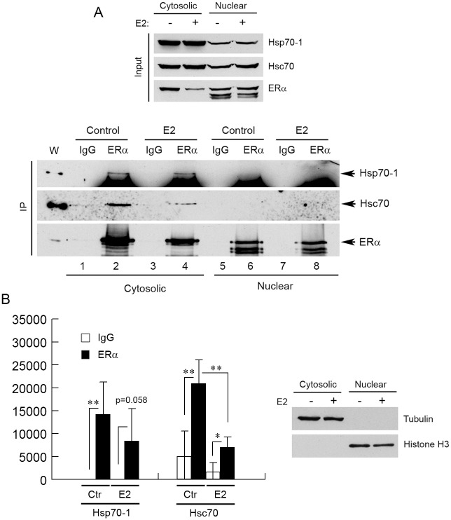
Fig 5. ERα interacts with Hsp70-1 and Hsc70 in the cytoplasm under conditions of hormone starvation/stimulation.
(A) The MCF7 cells were cultured under hormone starvation conditions for 3–4 days and then treated with either 100 nM E2 or ethanol (control) for 24 h. The cytosolic and nuclear extracts of the treated cells were then immunoprecipitated by anti-ERα antibody or a control IgG, and the immunoprecipitated protein was analyzed by Western blotting with the indicated antibodies. (B) Left panel, quantification of Western blots. Only the Hsp70-1 and Hsc70 protein bands in the cytosolic fractions were quantified. Signal intensity values in the Western blot quantifications were arbitrary numbers obtained by analyzing the protein bands with ImageJ software. Values in the Western blot quantifications were the means ± S.D. of four separate sample preparations. Right panel, validation of the cytosolic and nuclear fractionations. Tubulin and histone H3 were used as markers for the cytosolic and nuclear fractions, respectively. W, whole cell lysate. Ctr, control. * and ** denote p < 0.05 and p < 0.01, respectively.

