Figure 1.

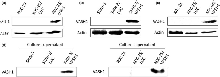
Establishment of soluble vascular endothelial growth factor receptor‐1 (sFlt‐1)‐ and vasohibin‐1 (VASH1)‐expressing cell lines. (a) Western blot with an anti‐vascular endothelial growth factor receptor‐1 antibody. The expression of the sFlt‐1 protein was only observed in KOC‐2S/sFlt‐1 ovarian cancer cells. (b, c) Western blots with an anti‐VASH1 antibody. The expression of the VASH1 protein was only observed in SHIN‐3/VASH1 (b) and KOC‐2S/VASH1 (c) ovarian cancer cells. Actin was used as a positive control. (d) Western blot of the culture supernatant, using an anti‐VASH1 antibody. The expression of the VASH1 protein in the culture supernatant was only observed for SHIN‐3/VASH1 and KOC‐2S/VASH1.
