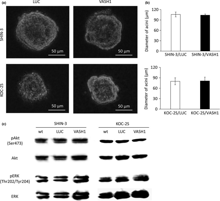
Figure 2.

Influence of vasohibin‐1 (VASH1) on cell growth and the cell signaling pathway. (a) In vitro cell growth in 3D cultures of SHIN‐3/LUC and SHIN‐3/VASH1 cells, and of KOC‐2S/LUC and KOC‐2S/VASH1 cells, as observed under a light microscope. (b) No significant differences were noted in the diameter of acini between the two groups for either cell line. (c) Western blots with anti‐protein kinase B (Akt), anti‐pAkt (Ser473), anti‐ERK, and anti‐pERK (Thr202/Tyr204) antibodies. No significant difference was observed in the phosphorylated Akt or ERK levels in either of the cell lines.
