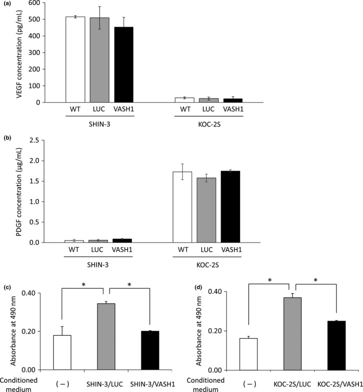
Figure 3.

Vascular endothelial growth factor (VEGF) and platelet‐derived growth factor (PDGF) concentrations in the supernatants from SHIN‐3 and KOC‐2S cultures, and HUVEC counts after the addition of the culture supernatant. (a) VEGF concentrations in the supernatants from SHIN‐3 and KOC‐2S cultures. Wild‐type SHIN‐3 cells were VEGF hypersecretory (515 ± 7 pg/mL), whereas WT KOC‐2S cells were not (28 ± 6 pg/mL). No significant difference was observed in the level of VEGF secretion between vasohibin‐1 (VASH1)‐transfected SHIN‐3 or KOC‐2S cell lines and WT or LUC‐transfected cells in each cell line. (b) PDGF concentrations in supernatants from SHIN‐3 and KOC‐2S cultures. Wild‐type SHIN‐3 cells were PDGF hyposecretory (0.049 ± 0.025 μg/mL), whereas WT KOC‐2S cells were PDGF hypersecretory (1.728 ± 0.192 μg/mL). (c) Effects of VASH1, secreted by SHIN‐3/VASH1 and KOC‐2S/VASH1 cells, on HUVECs. Absorbance was measured in order to obtain a relative value for the cell counts. The mean absorbance value was significantly higher for the group cultured with SHIN‐3/LUC culture supernatant (CS) than for the group without, but was significantly lower in the group cultured with SHIN‐3/VASH1 CS than the group with the SHIN‐3/LUC CS (*P < 0.01). (d) The absorbance value for the group cultured with KOC‐2S/LUC CS was significantly higher than for the group without, but was significantly lower for the group cultured with KOC2S/VASH1 CS than that with the KOC‐2S/LUC CS (*P < 0.01).
