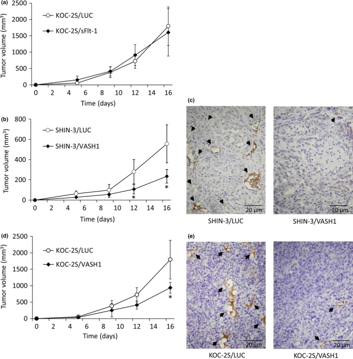
Figure 4.

Inhibition of tumor growth and vascularization by soluble vascular endothelial growth factor receptor‐1 (sFlt‐1) and vasohibin‐1 (VASH1). (a) Growth curves of s.c. KOC‐2S/LUC and KOC‐2S/sFlt‐1 tumors. No significant differences in tumor growth were noted between the KOC‐2S/LUC and KOC‐2S/sFlt‐1 groups. (b) Growth curves of s.c. SHIN‐3/LUC and SHIN‐3/VASH1 tumors. The tumor volume 12 days after inoculation and thereafter was significantly smaller in the SHIN‐3/VASH1 group compared to the SHIN‐3/LUC group. (c) CD31 immunohistochemical staining, developed with diaminobenzidine, of new blood vessels (black arrows) in SHIN‐3/LUC and SHIN‐3/VASH1 tumor tissues. (d) Growth curves of s.c. KOC‐2S/LUC and KOC‐2S/VASH1 tumors. Tumor volume 16 days after inoculation was significantly lower in the KOC‐2S/VASH1 group than the KOC‐2S/LUC group. (e) CD31 immunohistochemical staining, developed with diaminobenzidine, of new blood vessels (black arrows) in KOC‐2S/LUC and KOC‐2S/VASH1 tumor tissues. Data points for the graphs are represented as mean ± SD. *P < 0.01.
