Figure 5.

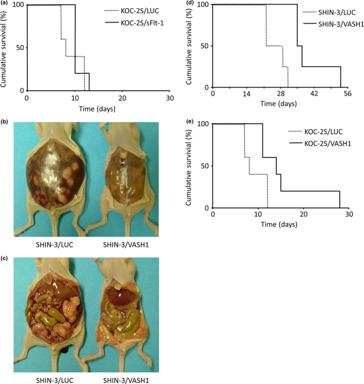
Inhibition of peritoneal dissemination by soluble vascular endothelial growth factor receptor‐1 (sFlt‐1) and vasohibin‐1 (VASH1). (a) Survival curves for mice i.p. transplanted with either KOC‐2S/LUC or KOC‐2S/sFlt‐1 cells. No significant difference was noted between the two groups. (b, c) Representative images of the laparotomic findings, carried out 21 days after i.p. SHIN‐3/LUC or SHIN‐3/VASH1 transplantation into mice. A large volume of ascites (b, left) and many peritoneal disseminated lesions (c, left) were observed in SHIN‐3/LUC‐transplanted mice. In contrast, no ascites (b, right) or peritoneal dissemination (c, right) was noted in SHIN‐3/VASH1‐transplanted mice. (d) Survival curves of mice i.p. transplanted with SHIN‐3/LUC or SHIN‐3/VASH1 cells. The survival time was significantly longer in the SHIN‐3/VASH1 group than in the SHIN‐3/LUC group (P < 0.01). (e) Survival curves of mice i.p. transplanted with KOC‐2S/LUC or KOC‐2S/VASH1 cells. The survival time was significantly longer in the KOC‐2S/VASH1 group than in the KOC‐2S/LUC group (P < 0.01).
