
Figure 5.
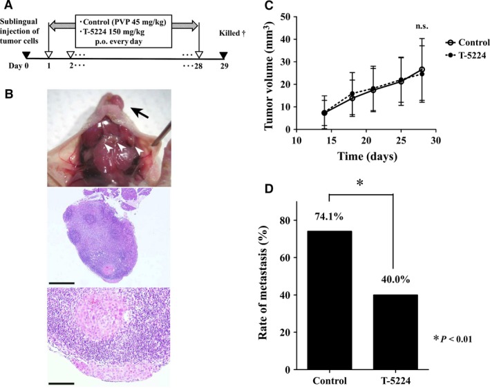
Protocol and results of in vivo study using an orthotopic model of head and neck squamous cell carcinoma. (A) Timeline of in vivo study. After HSC‐3‐M3 cells were injected into the tongue, vehicle or T‐5224 were given every day for 4 weeks. PVP, polyvinylpyrrolidone. †All mice were sacrificed on the 29th day after tumor inoculation into the tongue. (B) Primary lesion of tumor (arrow) and cervical lymph nodes (arrowhead) were observed. Lymph nodes were resected, and metastasis was assessed by H&E staining. Scale bar = 500 μm (middle image) and 100 μm (bottom image). (C) Tumor growth in the primary lesion. Tumors size did not differ significantly between the groups. Data represent mean ± SD. (D) The rate of lymph node metastasis was significantly lower in the T‐5224‐treated group than in the control group. *Statistically significant difference from the control is shown (P < 0.01).