Figure 2.

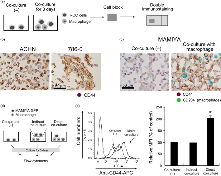
CD44 expression in cultured renal cell carcinoma (RCC) cell lines. (a) Cultured cells were prepared as cell‐block specimens and double immunostaining was performed. (b) CD44 expression on ACHN and 786‐O cells was evaluated by immunostaining. (c) Following co‐culture with macrophages for 3 days, CD44 expression in MAMIYA cells was evaluated by double immunostaining. Anti‐CD204 antibody was used to label macrophages (green), and we evaluated CD44 expression (brown) on CD204− cancer cells. (d) Following co‐culture with macrophages for 3 days, CD44s expression in RCC cell lines was evaluated by flow cytometry. (e) Following flow cytometry analysis, the mean fluorescence intensity (MFI) of CD44 was evaluated and statistically analyzed.
