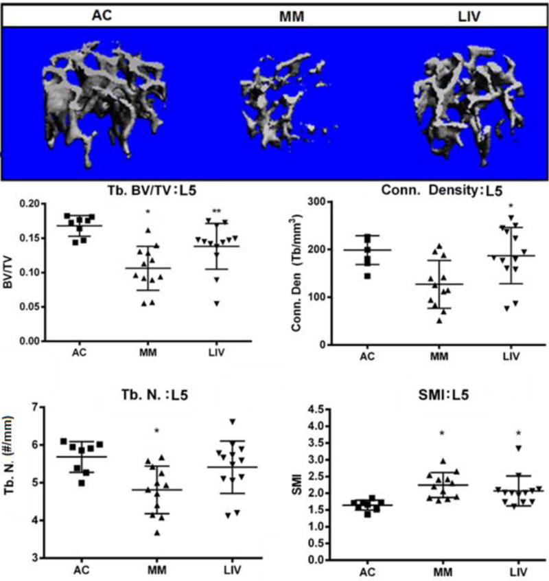
Figure 5.

Representative micro-CT (voxel resolution = 10μm) analysis of the L5 region of the axial skeleton. Trabecular bone was digitally isolated from the cortical shell of the vertebral body in order to quantify bone quantity and quality. BV/TV in MM mice were 35% (p<0.001) lower than in AC, LIV mice had 17% (NSD) less bone than AC, a measure that was 27% (p<0.05) greater as compared to MM. Trabecular connectivity density in MM mice was 36% (p<0.05) lower than in AC, whereas LIV mice were only 6% (NSD) less than in AC, a measure that was 47% (p<0.05) greater than those of MM. The number of trabeculae in the L5 of MM had decreased by 16% (p<0.05) versus AC while decreasing by just 5% (NSD) in LIV in comparison to AC, a mark that was 13% (NSD) greater than MM. The structure model index (SMI) indicated that trabecular bone was more rod-like in MM and LIV than AC, as this index was 37% (p<0.01) greater in MM than AC, but just 31% (p<0.05) greater in LIV than AC.
