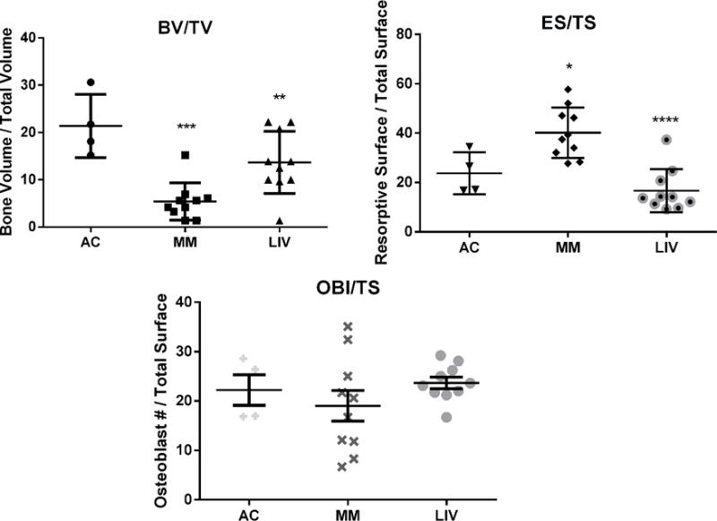
Figure 7.

Static histomorphometry was performed on histological sections of distal femora to discern the degree to which LIV effected resorption or formation. (Top Left) BV/TV was significantly reduced (p<0.0001) in MM from AC, a mark which improved by 60% (p<0.001) in the LIV-treated animals, all of which reflected data quantified via micro-CT analyses. (Top Right) As a measure of degree of bone resorption, resorptive surface per total surface was 70% (p<0.05) and 29% (NSD) greater in MM and LIV, respectively, as compared to AC. Introduction of LIV appears to have mitigated the degree of resorption by 58% (p<0.0001) than in myeloma animals denied LIV treatment. However, as an indication of bone formation, (Bottom) osteoblast counts in the same region (OBl/TS) were 14% (NSD) lower in MM as compared to AC mice. LIV appears to encourage a modest 24% (NSD) increase from MM-only mice. Overall, this indicates that a more anti-resorptive mechanism on trabecular surfaces was responsible for mitigating the osteolysis.
