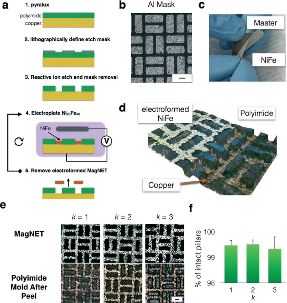
Fig. 2.
Master/replica fabrication of MagNET. a. Step by step fabrication of the master and subsequent replicas of MagNET. b. A micrograph of the lithographically defined aluminum mask. Scale bar 30 µm. c. Photograph of a replica MagNET being mechanically removed from its Master. d. Three dimensional optical micrograph of MagNET. In the region to the left, a MagNET has been electroformed. In the region to the right, the MagNET has been removed and the polyimide and copper master can be seen. e. Micrographs of the master and replica MagNETs after k replications. Scale bar: 30 µm. f. The fraction of damaged pillars was quantified after each replication, and there was no statistically significant change observed. (P ≫ 0.05). Error bars indicate standard error from the ratio of intact pores to total number of pores of different regions from the same device.

