
Figure 1.
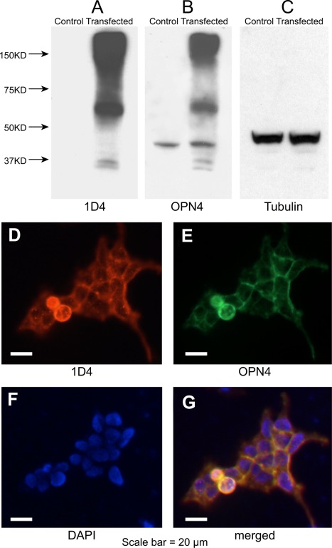
Immunocharacterization of the melanopsin antibody. Top: total proteins extracted from transfected T‐Rex cells probed with anti‐1D4 (A), antimelanopsin (B), and antitubulin (C) antibodies. Bands were detected by chemiluminescence corresponding to the expected sizes of 1D4‐tagged melanopsin fusion protein monomers (54 kDa) and oligomers (over 150 kDa). Bottom: transfected T‐Rex cells expressing 1D4‐tagged melanopsin fusion proteins were immunolabeled with anti‐1D4 (D) and antimelanopsin (E) antibodies. F: DAPI staining. G: Merged image indicates colocalization. Scale bars = 20 µm.