
Figure 10.
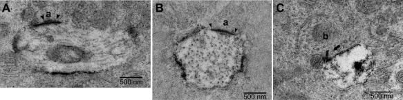
Synaptic contacts in the outer IPL in EM sections of melanopsin‐labeled macaque retina. A: A labeled dendrite cut in longitudinal section receives a synapse from an amacrine cell (a). The amacrine cell is relatively electron lucent and displays clear presynaptic specialization (arrowheads). B: A labeled dendrite cut in cross‐section receives a synapse from an amacrine cell (a). Because of the dense labeling of the ganglion cell membrane, synapses were detected based on presynaptic specializations (arrowheads). C: A labeled ganglion cell dendrite cut in cross‐section receives a synapse from a cone bipolar cell axon (b) at a ribbon synapse (arrow). The other member of the dyad is an amacrine cell process. Scale bars = 500 µm.