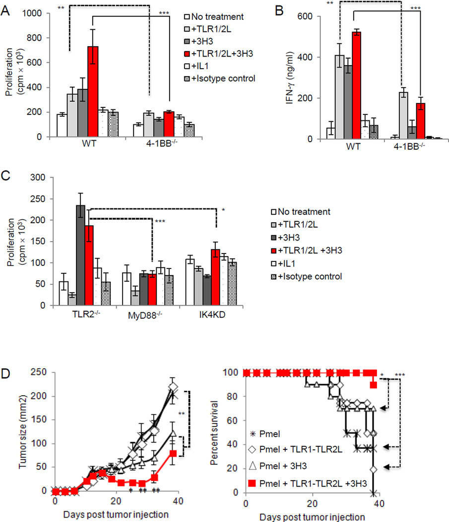
Figure 5. Combining TLR1–TLR2 ligand and 4-1BB agonistic antibody increases T-cell proliferation and improves anti-tumor activity against melanoma in mice.
A and B, Purified WT and 4-1BB−/−, or (C),TLR2−/−, MyD88−/− and IRAK4 kinase dead CD8+ T cells were activated by CD3ε Ab-coated plates in the presence of TLR1–TLR2L, 3H3, TLR1–TLR2L and 3H3, IL1, or isotype antibody. Proliferation (±s.d.) was assessed by measuring 3H-thymidine uptake, whereas IFN-γ production was measured by ELISA 48 hours after activation. Student t test, ** P <0.01, *** P <0.001. Data from (A–C) are representative of three independent experiments. D, B16-F1 melanoma cells were injected subcutaneously into C57B6 mice. When palpable tumors were detected mice were irradiated and injected intravenously with 5 × 10⁶–7 × 10⁶ activated pmel T cells that had been exposed to TLR1–TLR2 ligand 24 hours before injection. TLR1–TLR2 ligand and 3H3 were injected intraperitoneally. Error bars represent standard error from the mean of 10 mice per group. Statistics were generates by repeated measures ANOVA-Tukey post test, *P <0.05, **P <0.01, ***P <0.001. Survival statistics were generated by log rank test. Student t test was conducted between 3H3 and TLR1–TLR2L + 3H3. ‡P <0.05, ‡‡P <0.01.

