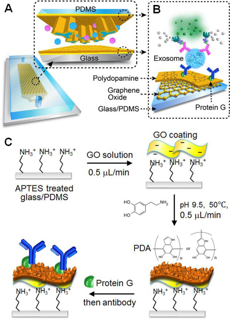
Figure 1.
The nano-interfaced microfluidic exosome platform (nano-IMEX). (A) Schematic of a single-channel PDMS/glass device, with the exploded-view highlighting the coated PDMS chip containing an array of Y-shaped microposts. (B) Surface of the channel and microposts coated with graphene oxide (GO) and polydopamine (PDA) as a nanostructured interface for the sandwich ELISA of exosomes with enzymatic fluorescence signal amplification. (C) The procedure for surface functionalization of the microfluidic chips.

