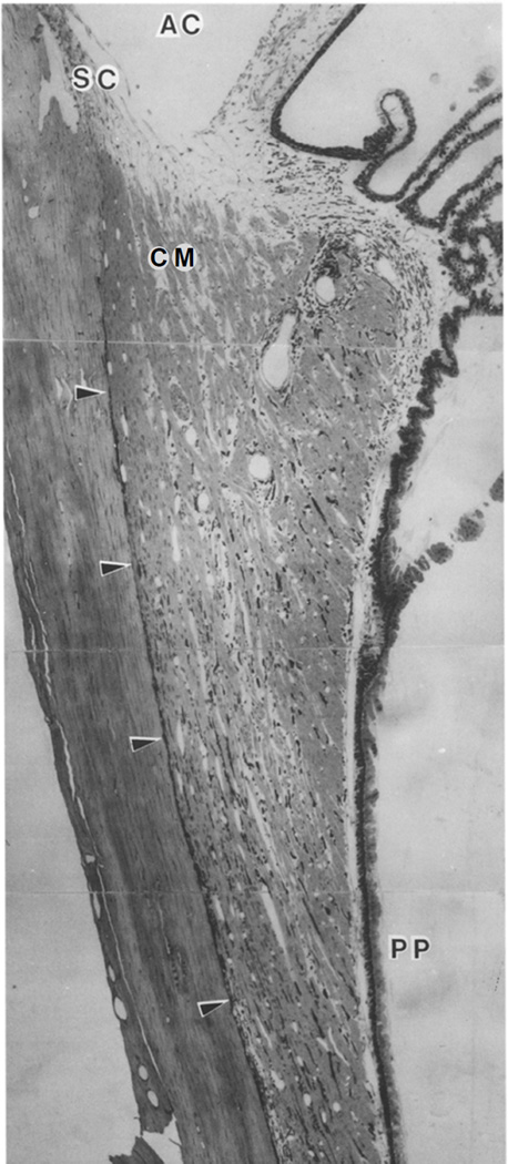
Figure 1.
Meridional section through the uveal tract of a Macaca fascicularis . Arrowheads show supraciliary and suprachoroidal space. Unconventional outflow passes from the anterior chamber (AC), through the most posterior aspects of the uveal meshwork, enters the open spaces between longitudinal aspects of the ciliary muscle (CM) and then enters the suprachoroidal space. SC -- Schlemm’s canal; PP -- pars plana. (Wood et al. 1990)
[Permission Needed]

