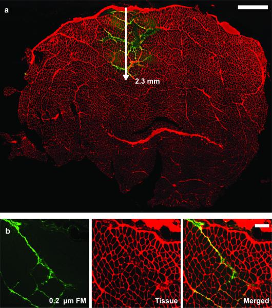
Figure 2.
a) Representative TA muscle cross-section at the injection site with the fluorescent microspheres (FM) (green) in the extracellular space (red). Fluorescent microspheres dispersed at a depth of 2.3 mm into the tissue, remaining in the interstitial space. Scale is 1 mm. b) Transverse section of TA taken about 5 mm away from the injection site. The 0.2 μm fluorescent microspheres (green) stay within the interstitial space (red) and do not penetrate the muscle fiber. Scale bar is 200 μm.

