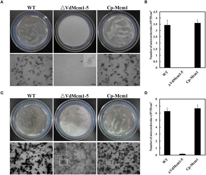
Figure 5.
Loss of VdMcm1 causes defects in microsclerotia formation. (A) Microsclerotia formation on BM plates with 105 conidia spreading over the cellulose membrane after 6 days of growth. The enlarged view shows the unmelanized and swollen hyphae of ΔVdMcm1-5. The scale bars represent 100 μm. (B) The histogram represents the number of microsclerotia formed by the wild-type strain, ΔVdMcm1-5, and complemented strain on the cellulose membrane (φ = 80 mm) after 6 days of growth. (C) Microsclerotia formation on BM plates after 8 days of incubation. The enlarged view showed the microsclerotia of ΔVdMcm1-5. The scale bars represent 100 μm. (D) The histogram represented the number of microsclerotia formed by wild-type strain, ΔVdMcm1-5, and complemented strain on the cellulose membrane (φ = 80 mm) after 8 days of incubation. The error bars represent standard deviations. The experiments were performed in triplicate.

