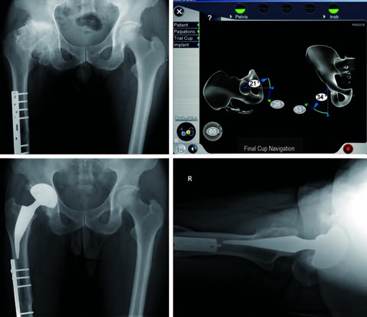
Fig. 3.
A 62-year-old man with a deformed acetabulum in the right hip. (A) Preoperative anteroposterior hip radiograph. (B) Intraoperative navigation data. The inclination and anteversion values displayed in the fields with gray and white backgrounds are for the initial situation and for final cup implantation, respectively. (C) Anteroposterior hip radiograph at 1-year follow-up. (D) Translateral radiographs of the right hip showing that the components are stable with no evidence of dislocation.

